Mouse EPO/Erythropoietin enzyme-linked immunoassay kit
| Specification | 96 Test |
|---|---|
| Sensitivity | 0.07 pg/ml (10 μl) |
| Standard Curve Range | 2.74~2000 pg/ml |
| Standard Curve Gradient | 7 Points/3 Folds |
| Number of Incubations | 2 |
| Detectable sample | Liquid phase sample of soluble substances. For example: serum, plasma, cell culture supernatant, tissue grinding liquid, etc. |
| Sample Volume | 10 μl |
| Type | Fully Ready-to-Use |
| Operation Duration | 120min |
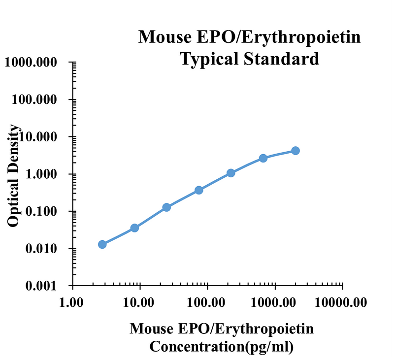
| pg/ml | O.D. | Average | Corrected | |
|---|---|---|---|---|
| 0.00 | 0.0178 | 0.0135 | 0.0157 | |
| 2.74 | 0.0304 | 0.0264 | 0.0284 | 0.0128 |
| 8.23 | 0.0474 | 0.0548 | 0.0511 | 0.0355 |
| 24.69 | 0.1433 | 0.1391 | 0.1412 | 0.1256 |
| 74.07 | 0.4046 | 0.3610 | 0.3828 | 0.3672 |
| 222.22 | 1.1100 | 1.0450 | 1.0775 | 1.0619 |
| 666.67 | 2.6610 | 2.6220 | 2.6415 | 2.6259 |
| 2000.00 | 4.1867 | 4.2902 | 4.2385 | 4.2228 |
Precision
| Intra-assay Precision | Inter-assay Precision | |||||
| Sample Number | S1 | S2 | S3 | S1 | S2 | S3 |
| 22 | 22 | 22 | 6 | 6 | 6 | |
| Average(pg/ml) | 32.9 | 169.3 | 528.9 | 33.9 | 176.3 | 571.3 |
| Standard Deviation | 0.9 | 3.3 | 20.1 | 0.9 | 9.0 | 27.4 |
| Coefficient of Variation(%) | 2.9 | 1.9 | 3.8 | 2.7 | 5.1 | 4.8 |
Intra-assay Precision (Precision within an assay) Three samples of known concentration were tested Twenty-two times on one plate to assess intra-assay precision.
Inter-assay Precision (Precision between assays) Three samples of known concentration were tested six times on one plate to assess intra-assay precision.
Spike Recovery
The spike recovery was evaluated by spiking 3 levels of mouse EPO/Erythropoietin into health mouse serum sample. The un-spiked serum was used as blank in this experiment.
The recovery ranged from 74% to 123% with an overall mean recovery of 102%.
Sample Values
| Sample Matrix | Sample Evaluated | Range (pg/ml) | Detectable (%) | Mean of Detectable (pg/ml) |
|---|---|---|---|---|
| Serum | 30 | 6.43-488.96 | 100 | 71.25 |
Serum/Plasma – Thirty samples from apparently healthy mice were evaluated for the presence of EPO/Erythropoietin in this assay. No medical histories were available for the donors. n.d. = non-detectable. Samples measured below the sensitivity are considered to be non-detectable.
Product Data Sheet
Background: EPO/Erythropoietin
Erythropoietin (Epo) is a 34-39 kDa secreted glycoprotein that is a member of the type I cytokine superfamily. The mouse Epo gene encodes a 192 amino acid (aa) residue precursor that contains a 26 aa signal peptide and a 166 aa mature protein containing three potential N-linked glycosylation sites. Mouse Epo lacks the O-linked glycosylation site found in human Epo. Although carbohydrate chains are not required for in vitro receptor binding, they are required for in vivo Epo bioactivity. Depending on the cell source, different Epo isoforms are produced that differ in their glycan compositions and sialic acid contents. Mature mouse and rat Epo share 94% aa sequence identity. They also share from 80%-82% aa identity with mature human, porcine, rhesus monkey and feline Epo. Epo is primarily produced by cells in the kidney (interstitial peritubular renal fibroblasts) and liver (hepatocytes and Ito cells), where its production is up-regulated by hypoxia. Other tissues and cells, including neural tissues (astrocytes and neurons), testis (Sertoli cells), uterus, placenta, and erythroid progenitors, have also been shown to produce Epo.
Epo is best known for its role in red blood cell formation. While Epo is not a lineage commitment factor, it inhibits apoptosis and induces burst forming unit-erythroid (BFU-E) differentiation into colony forming unit-erythroid (CFU-E), and the subsequent proliferation and maturation of CFU-E into early normoblasts. Apart from its role in erythropoiesis, Epo also acts on various non-hematopoietic cells to function as a viability and proliferation factor. Epo can stimulate myoblast proliferation while suppressing its differentiation, resulting in the expansion of the progenitor cell population. Epo is a tissueprotective factor that protects against ischemic and toxic injuries to neuronal, cardiovascular and renal tissues. Epo has also been shown to promote angiogenesis in various physiologic and pathologic conditions.
Epo binds and signals via the high-affinity preformed homodimeric Epo receptor (Epo R) that is composed of two Epo R subunits. Each Epo R subunit is a type I transmembrane glycoprotein that belongs to the type I cytokine receptor superfamily. Its extracellular domain contains the characteristic two fibronectin type III domains and a WSxWS motif near the plasma membrane. Binding of Epo to the Epo R homodimer results in conformational change and phosphorylation and activation of the non-receptor protein kinase JAK2, which activates the downstream signaling cascade. An alternative Epo heteromeric receptor complex that transduces cell-protective signals and containing the beta common receptor ( beta CR) subunit in addition to the Epo R subunit has been described. beta CR also belongs to the type I cytokine receptor superfamily and is a subunit that is shared by the heteromeric IL-3, IL-5 and GM-CSF receptor complexes. Epo binds with lower affinity to the heteromeric receptor consisting of a Epo R subunit and a beta CR homodimer.